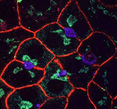
| WIF-B cells are
highly-polarized. Triple-immunofluorescence labeling of apical
(blue), basolateral (red) and Golgi (green) proteins in fully polarized
WIF-B cells. Fixed and permeabilized cells were incubated with
anti-HA4, anti-HA321 and anti-albumin antibodies of three different
species (mouse, rabbit and guinea pig, respectively) followed by
appropriate fluorescently-labeled secondary antibodies. The fluorescence
image of each label was recorded on a separate channel of a confocal laser
scanning microscope though three ~1 um optical sections starting ~6 um
from the bottom. The images were merged into one image and
pseudocolored. The apical protein, HA/4cell-CAM105/ ectoATPase, is
localized to the membrane lining the dilated spaces, whereas the HA321/BEN
protein is found along the basolateral surface and albumin is concentrated
in the Golgi. The nuclear-Golgi-apical axis is clearly evident in
these cells. |
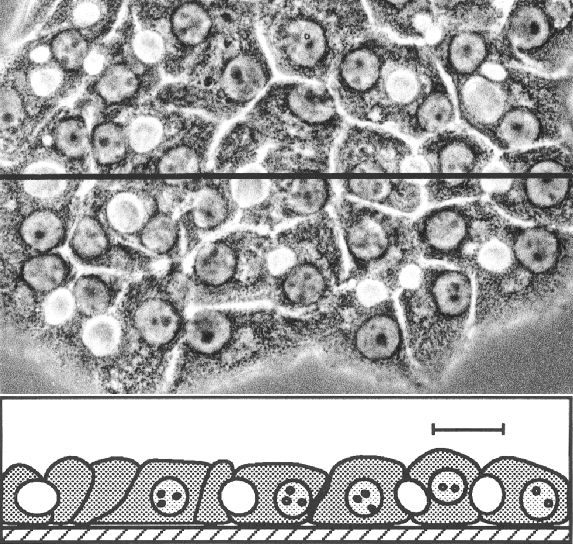
Phase contrast
morphology of living WIF-B cells in culture. Top:
WIF-B cells were plated onto tissue culture plastic and grown for >10
days. The cells have a polygonal appearance and are closely
packed. Phase-lucent "blisters" between neighboring
cells are clearly evident and correspond to the "bilie
canalicular" or apical space. Bottom: A cross-sectional
"view" of the cell monolayer taken along the line indicated in
the upper panel. The phase-lucent spaces are closed off from both
the substrate and the overlaying medium.
|
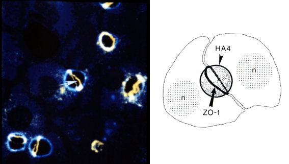
ZO-1 marks the borders
between apical and basolateral domains. WIF-B cells were double
labeled for the tight junction-associated protein ZO-1 and the apical
marker HA4. Schematic drawing of the BC which is marked by an arrow
and/or arrowhead (n=nucleus of adjacent cell). ZO-1 (arrows)
was visualized with rat mAb followed by goat FITC-IgG, and HA4 (arrowheads
was stained with rabbit pAb followed by goat Texas red-IgG. The
image of ZO-1 is a compiled z-series taken in 1 um steps, and that of HA4
is a 1 um optical x-y section. Both images were merged to generate
the image in C to illustrate the three dimensional BC architecture.
The belt-like presence of ZO-1 around BC between the participating cells
suggests the existence of tight-junctional structures separating apical
and basolateral domains. One can see an intercellular "vacuolar
compartment" (v) which is positive for HA4 but has no ZO-1
staining. Bar, 10 um. See Ihrke, et all (1993), JCB, 1761-1775
for details. |