Research Images
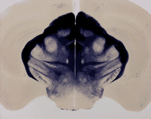 Coronal section of an adult mouse brain at the level of the thalamus shows the axons, dendrites, and cell bodies of Brn3b expressing neurons. This mouse carries an alkaline phosphatase reporter knocked into the Brn3b locus, and the pattern of alkaline phosphatase localization was revealed histochemically.
![Coronal section of an adult female mouse brain showing the distribution of neurons expressing each of the X-chromosomes. [As a consequence of X-chromosome inactivation, only one X-chromosome is expressed in each cell.] One X-chromosome expresses a nuclear localized GFP and the other X-chromosome expresses a nuclear localized tdTomato. Substantial left-right asymmetry is apparent.](images/X-inactivation_in_the_mouse_brain.jpg)
